Label the transmission electron micrograph of the mitochondrion – Labeling the transmission electron micrograph of the mitochondrion is a crucial step in understanding its intricate ultrastructure and function. This detailed guide provides a comprehensive overview of the mitochondrial morphology, guiding readers through the identification of key structures and their significance in cellular processes.
Mitochondrial Ultrastructure

Mitochondria are organelles found in eukaryotic cells that are responsible for cellular respiration. They have a distinct structure that enables them to carry out their functions efficiently.
Overall Shape and Size, Label the transmission electron micrograph of the mitochondrion
Mitochondria are typically rod-shaped or oval and range in size from 0.5 to 1 micrometer in length. Their size and shape can vary depending on the cell type and its metabolic needs.
Mitochondrial Membranes
Mitochondria are enclosed by two membranes: an outer membrane and an inner membrane. The outer membrane is smooth, while the inner membrane is highly folded, forming structures called cristae.
- Outer Membrane:Permeable to small molecules and ions due to the presence of porins. It regulates the entry of molecules into the mitochondrion.
- Inner Membrane:Impermeable to most molecules. It contains proteins involved in oxidative phosphorylation, the process that generates ATP.
Matrix and Cristae
The space enclosed by the inner membrane is filled with a gel-like substance called the matrix. The matrix contains enzymes involved in the Krebs cycle, fatty acid oxidation, and other metabolic processes.
The cristae increase the surface area of the inner membrane, providing more space for the electron transport chain and ATP synthase, which are involved in oxidative phosphorylation.
Label the Transmission Electron Micrograph

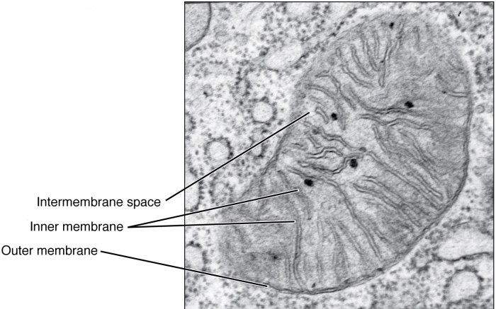
The transmission electron micrograph (TEM) image of the mitochondrion reveals its intricate structure.
Structures Labeled:
- Outer Membrane:Smooth, outer boundary of the mitochondrion.
- Inner Membrane:Highly folded, forming cristae.
- Matrix:Dense, gel-like substance within the inner membrane.
- Cristae:Folds of the inner membrane that increase its surface area.
- Ribosomes:Small, dark particles attached to the inner membrane or cristae, involved in mitochondrial protein synthesis.
Mitochondrial Functions

Mitochondria play crucial roles in cellular respiration and other cellular processes.
Cellular Respiration
Mitochondria are the primary sites of cellular respiration, the process that converts glucose into ATP, the energy currency of cells.
- Glycolysis:Occurs in the cytoplasm, breaking down glucose into pyruvate.
- Krebs Cycle (Citric Acid Cycle):Occurs in the mitochondrial matrix, further breaking down pyruvate to produce CO2 and energy carriers (NADH and FADH2).
- Oxidative Phosphorylation:Occurs on the inner mitochondrial membrane, using NADH and FADH2 to generate ATP.
Oxidative Phosphorylation
Oxidative phosphorylation is a multi-step process that generates most of the ATP in the cell.
- Electron Transport Chain:A series of proteins embedded in the inner membrane that transfer electrons from NADH and FADH2 to oxygen.
- ATP Synthase:A protein complex that uses the energy released from electron transfer to pump protons across the inner membrane, creating a proton gradient.
- ATP Production:As protons flow back through ATP synthase, they drive the synthesis of ATP from ADP.
Apoptosis
Mitochondria play a role in apoptosis, or programmed cell death.
- Cytochrome c Release:In response to apoptotic signals, mitochondria release cytochrome c into the cytoplasm.
- Caspase Activation:Cytochrome c triggers the activation of caspases, a family of enzymes that lead to cell disassembly.
Mitochondrial Disorders: Label The Transmission Electron Micrograph Of The Mitochondrion

Mitochondrial disorders are a group of genetic conditions that result from defects in mitochondrial function.
Common Disorders
- Mitochondrial Encephalopathy, Lactic Acidosis, and Stroke-like Episodes (MELAS):Characterized by seizures, strokes, and lactic acidosis.
- Leigh Syndrome:A severe neurodegenerative disorder that affects infants and young children.
- Kearns-Sayre Syndrome:A multi-system disorder that includes muscle weakness, heart problems, and vision loss.
Genetic Basis
Mitochondrial disorders are typically inherited from the mother, as mitochondria are inherited exclusively through the egg cell.
- Nuclear Gene Mutations:Mutations in nuclear genes that encode mitochondrial proteins can lead to mitochondrial dysfunction.
- Mitochondrial DNA Mutations:Mutations in the mitochondrial DNA itself can also cause mitochondrial disorders.
Challenges in Diagnosis and Treatment
Diagnosing mitochondrial disorders can be challenging due to their varied symptoms and overlapping presentations with other conditions.
Treatment options are limited, but may include:
- Symptomatic Treatment:Managing the symptoms of the disorder, such as seizures or muscle weakness.
- Mitochondrial Supplements:Providing nutrients that support mitochondrial function, such as coenzyme Q10 and L-carnitine.
- Gene Therapy:In some cases, gene therapy may be an option to correct mitochondrial DNA mutations.
Questions and Answers
What is the function of the mitochondrial matrix?
The mitochondrial matrix contains enzymes involved in various metabolic processes, including the citric acid cycle and fatty acid oxidation.
How are mitochondrial disorders inherited?
Mitochondrial disorders can be inherited through both maternal and paternal lines due to the presence of mitochondrial DNA.